What It Is
Canalicular laceration repair is a delicate surgical procedure used to treat injuries to the canaliculus—tiny tear drainage channels in the inner eyelids. These ducts carry tears from the eyes to the nose. When they are cut or torn, patients may develop constant tearing (epiphora), infections, and eyelid deformities.
In Korea, this surgery is performed by oculoplastic and reconstructive eye surgeons using advanced microsurgical instruments and silicone stenting to restore normal tear drainage and eyelid appearance.
Why It’s Done
Repair is performed when:
- The tear duct (canaliculus) is injured due to trauma, cuts, or animal bites
- Patients experience persistent watery eyes because of poor drainage
- There is a risk of permanent scarring or deformity if left untreated
- Eyelid shape and tear function need to be preserved after an accident
The goal is to restore both function and aesthetics of the eyelid and tear system.
Alternatives
- Observation: Very small or superficial eyelid cuts not involving the tear duct may heal without surgery
- Temporary stenting: Used in partial or uncertain injuries to keep ducts open
- Medical therapy: Antibiotics and wound care for mild injuries or to prepare for surgery
For complete canalicular injuries, surgery is almost always necessary to restore tear function.
Preparation
Preparation for surgery typically includes:
- Consultation: With an oculoplastic surgeon to confirm canalicular injury
- Eye examination: Checking eyelid function and tear duct integrity
- Wound stabilization: Cleaning and antibiotics if the injury is recent
- Anesthesia planning: Local anesthesia with sedation for adults, general anesthesia for children or complex trauma
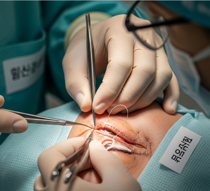
How It’s Done
The procedure takes about 1–2 hours.
- The surgeon identifies the cut canaliculus under magnification
- A silicone stent or tube is passed through the tear duct to maintain an open channel
- The lacerated canaliculus is repaired with fine sutures under a microscope
- The eyelid tissue is carefully reconstructed to restore natural contour
- The stent remains in place for several weeks to months until healing is complete
Recovery
- First week: Swelling, bruising, and mild irritation are common
- Activity: Avoid eye rubbing or heavy activity for a few weeks
- Follow-ups: To ensure the stent is in position and the canaliculus is healing
- Stent removal: Usually after 3–6 months
- Final results: Most patients regain normal tear function and eyelid shape
Possible Complications
- Infection or delayed wound healing
- Stent displacement or blockage
- Persistent tearing if the canaliculus does not heal properly
- Scar tissue or eyelid deformity
- Rare need for revision surgery
Treatment Options in Korea
Diagnosis
- Detailed examination of the eyelid and tear duct
- Probing of tear drainage channels to confirm injury
Medical Treatments
- Antibiotics and lubricating drops after surgery
- Pain management and scar ointments
Surgical or Advanced Therapies
- Microsurgical canalicular repair: With silicone stenting (monocanalicular or bicanalicular)
- Mini-Monoka stent technique: Widely used in Korea for less invasive repair
- Complex reconstruction: For severe trauma involving both eyelids and ducts
Rehabilitation and Support
- Scar management with creams or laser if needed
- Long-term monitoring of tear drainage
- International patient services including interpreters, hospital coordination, and telemedicine follow-up