What it is
An MRI (Magnetic Resonance Imaging) scan is a non-invasive imaging technique that uses strong magnetic fields and radio waves to create detailed images of internal organs, tissues, and structures.
Key points:
- Provides high-resolution images of the brain, spine, joints, abdomen, heart, and blood vessels.
- Does not use ionizing radiation (unlike X-rays or CT scans).
- Useful for diagnosing conditions, planning treatment, and monitoring disease progression.
- Often combined with contrast agents to enhance visibility of certain tissues or blood vessels.
Why it’s done
MRI scans are performed to:
- Diagnose diseases or injuries: Tumors, strokes, spinal cord issues, joint injuries, heart conditions.
- Monitor chronic conditions: Multiple sclerosis, cancer, or liver disease.
- Guide treatment: Pre-surgical planning, biopsy guidance, or radiation therapy targeting.
- Evaluate unexplained symptoms: Chronic pain, neurological deficits, or internal organ problems.
Note: MRI is especially valuable for soft tissue imaging, where other modalities may be less effective.
Alternatives
Other imaging options include:
- CT scan (Computed Tomography): Uses X-rays; better for bone injuries and acute bleeding.
- Ultrasound: Useful for abdominal organs, vessels, and pregnancy.
- X-rays: Best for bone fractures or lung imaging.
- PET scan: Often used for cancer staging or metabolic studies.
Important: MRI is preferred when high-resolution soft tissue detail is required.
Preparation
Preparation for an MRI scan depends on the type of study:
- Medical history: Inform staff of metal implants, pacemakers, or prior surgeries.
- Fasting: Required for some abdominal or pelvic scans.
- Clothing: Wear metal-free clothing; remove jewelry, watches, and piercings.
- Contrast considerations: Inform the technician of allergies or kidney problems if contrast is used.
Patient instructions:
- Stay still during the scan to ensure clear images.
- Follow any breathing instructions provided by staff.
- Ask about sedation options if claustrophobic.
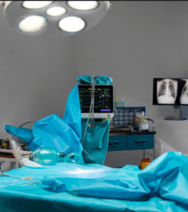
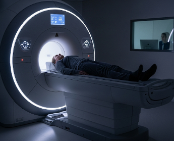
How it’s done
MRI scanning is performed in a specialized imaging room:
- Positioning: Patient lies on a motorized table that slides into the MRI machine.
- Coils placement: Special devices (“coils”) may be placed around the body part being imaged.
- Scanning: The machine generates magnetic fields and radio waves; patients hear loud knocking noises during the scan.
- Contrast injection (if required): Administered intravenously to highlight certain tissues.
- Duration: Typically 15–90 minutes, depending on the area being imaged.
Note: Patients must remain still throughout to avoid blurring.
Recovery / Post-Scan Considerations
MRI scans are non-invasive and require minimal recovery:
- No downtime: Patients can resume normal activities immediately.
- Hydration: Recommended after contrast-enhanced scans to help flush the agent.
- Results: Typically reviewed by a radiologist and sent to the referring doctor within hours to a few days.
- Follow-up: Additional imaging may be ordered if abnormalities are detected.
Benefits:
- Provides precise anatomical detail for diagnosis and treatment planning.
- Non-invasive and safe for repeated use.
- Helps in early detection and management of serious conditions.
Complications / Risks
MRI scans are generally safe, but potential risks include:
- Claustrophobia: Anxiety due to enclosed space.
- Noise discomfort: Loud knocking sounds during scanning.
- Contrast reactions: Rare allergic reactions to gadolinium-based agents.
- Implant interference: Patients with pacemakers, certain implants, or metal fragments may be at risk.
Prevention / Management:
- Screen for metallic implants or devices before scanning.
- Provide ear protection to reduce noise discomfort.
- Sedation can be offered for anxiety or claustrophobia.
- Monitor patients for contrast-related adverse reactions.
Treatment Options in Korea
MRI scanning is widely available in hospitals, specialized imaging centers, and clinics across Korea:
Key features:
- High-resolution 1.5T and 3T MRI machines for detailed imaging.
- Experienced radiologists and technicians ensure accurate imaging and reporting.
- Available for a wide range of diagnostic and treatment planning purposes.
- Facilities provide contrast-enhanced scans, functional MRI, and cardiac MRI.
- Supports early diagnosis, treatment monitoring, and improved patient outcomes.
Summary: MRI scanning in Korea is a safe, non-invasive, and highly effective imaging tool. With advanced equipment, expert staff, and comprehensive imaging protocols, patients benefit from accurate diagnosis, early treatment planning, and improved management of complex conditions.