Overview
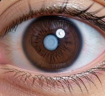
Leukocoria is a medical term for a white reflection or white pupil, often noticeable in photographs or in dim light. It is sometimes called a “cat’s eye reflex” and can affect one or both eyes. Leukocoria is not a disease itself but a sign of an underlying eye condition, which can range from benign to potentially vision-threatening or life-threatening.
In Korea, ophthalmology clinics and specialized pediatric eye centers provide advanced diagnostic services such as retinal imaging, ultrasonography, and genetic testing to identify the underlying cause of leukocoria. Early detection and treatment are essential to preserve vision and, in some cases, save life.
Key Facts
- ➔ Leukocoria manifests as a white or yellowish reflection in the pupil.
- ➔ It can be detected in photographs using flash or during routine eye examinations.
- ➔ Causes may include retinoblastoma, congenital cataract, Coats’ disease, or retinal detachment.
- ➔ Prompt evaluation is crucial, as some causes like retinoblastoma are potentially life-threatening.
- ➔ Treatment and prognosis vary depending on the specific underlying condition.
What is Leukocoria?
Leukocoria is not a disease itself, but a clinical sign indicating an abnormality in the retina or lens that prevents the normal red reflex seen in healthy eyes.
- ➔ Red reflex: Normally, light reflects from the retina, producing a reddish glow in photographs.
- ➔ White reflex: When light reflects abnormally, it appears white, signaling a structural or retinal problem.
- ➔ Clinical importance: Leukocoria may be the first sign of serious eye conditions, especially in infants and young children.
- ➔ Detection: Can be identified during routine eye exams, photographs, or parental observation.
What Symptoms Are Related To
Leukocoria may occur with other ocular symptoms depending on the underlying cause:
- ➔ Strabismus (misaligned eyes)
- ➔ Poor vision or visual impairment
- ➔ Eye redness or swelling
- ➔ Eye pain or sensitivity to light
- ➔ Nystagmus (involuntary eye movements)
- ➔ Differences in pupil size or shape
- ➔ Delayed visual development in infants
Recognizing these associated symptoms is vital for early intervention and management.
What Causes / Possible Causes
Leukocoria can result from a variety of eye disorders:
- ➔ Retinoblastoma: A rare but aggressive eye cancer, often affecting children under five.
- ➔ Congenital cataract: Clouding of the lens present at birth, affecting vision development.
- ➔ Coats’ disease: Abnormal retinal blood vessels causing retinal detachment and vision loss.
- ➔ Persistent fetal vasculature: A developmental eye abnormality present from birth.
- ➔ Retinal detachment or trauma: Can prevent normal red reflex and create white reflection.
- ➔ Toxocariasis or ocular infections: Parasitic or infectious causes leading to retinal lesions.
Early recognition of the cause is essential to prevent vision loss and, in the case of tumors, to save life.
When Should I See My Doctor
Immediate evaluation is necessary if leukocoria is observed:
- ➔ White reflection in the pupil noticed in photographs or during routine observation
- ➔ Strabismus or eye misalignment in infants or children
- ➔ Poor visual response or developmental delay
- ➔ Redness, swelling, or eye pain
- ➔ Family history of eye disorders or retinoblastoma
Prompt assessment by a pediatric ophthalmologist or eye specialist is critical for early diagnosis and effective treatment.
Care and Treatment
Treatment of leukocoria depends entirely on the underlying cause:
- ➔ Retinoblastoma: Treatment may include chemotherapy, laser therapy, cryotherapy, radiation, or surgical removal of the eye (enucleation) in severe cases.
- ➔ Congenital cataract: Surgical removal of the cataract, often followed by corrective lenses or intraocular lens implantation.
- ➔ Coats’ disease: Laser therapy or cryotherapy to repair retinal vessels and prevent retinal detachment.
- ➔ Persistent fetal vasculature: Surgical correction may be needed to restore normal eye anatomy.
- ➔ Retinal detachment: Surgical reattachment using vitrectomy, scleral buckling, or pneumatic retinopexy.
- ➔ Supportive care: Regular vision monitoring, corrective lenses, and rehabilitation if vision impairment persists.
Early treatment can restore or preserve vision, prevent complications, and in the case of retinoblastoma, save the child’s life.
Treatment Options in Korea
Korean hospitals provide comprehensive care for leukocoria:
- ➔ Advanced diagnostic imaging: Ultrasound, fundus photography, and MRI for precise assessment.
- ➔ Genetic testing: Especially important in retinoblastoma for family counseling and risk assessment.
- ➔ Surgical interventions: Pediatric ophthalmic surgeons perform cataract removal, retinal repairs, or enucleation when necessary.
- ➔ Laser and cryotherapy treatments: For vascular abnormalities like Coats’ disease.
- ➔ Chemotherapy and radiotherapy: For malignant tumors such as retinoblastoma.
- ➔ Multidisciplinary approach: Collaboration among ophthalmologists, pediatric oncologists, radiologists, and genetic counselors to provide personalized care.
Leading hospitals such as Seoul National University Hospital, Samsung Medical Center, and Asan Medical Center offer state-of-the-art pediatric ophthalmology services, ensuring early detection and effective treatment of leukocoria.
In Summary: Leukocoria is a white pupil reflex indicating an underlying eye abnormality, ranging from congenital cataracts to life-threatening tumors like retinoblastoma. Early evaluation, accurate diagnosis, and advanced treatment in Korea can preserve vision, restore eye function, and save lives.
- ➔ Key Takeaway: Any white reflection in a child’s pupil should prompt immediate medical evaluation.
- ➔ Action Point: Seek care from a pediatric ophthalmologist or specialized eye center for thorough assessment and management.