Overview
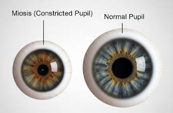
Eye miosis refers to the abnormal constriction of the pupils, resulting in smaller-than-normal pupil size. The pupil controls the amount of light entering the eye, and its size is normally regulated by the autonomic nervous system. Miosis can be physiological, drug-induced, or a sign of an underlying medical condition. In Korea, ophthalmology and neurology clinics provide comprehensive evaluation and management of miosis to ensure proper diagnosis and prevent complications related to vision or neurological health.
Highlights:
➤ Abnormally small pupils – Can affect light entry and vision
➤ Caused by drugs, neurological conditions, or ocular diseases
➤ May be an early sign of systemic or neurological disorders
Key Facts
➤ Normal pupil size: Typically 2–4 mm in bright light and 4–8 mm in dim light
➤ Prevalence: Miosis is less common than mydriasis (dilated pupils) but occurs in specific conditions or drug exposures
➤ Age affected: Can occur at any age depending on cause
➤ Gender: Both males and females can be affected
➤ Impact: May result in difficulty seeing in low light, eye strain, or indicate serious systemic conditions
What is Eye Miosis?
Miosis is defined as a persistent or excessive constriction of the pupils. The condition can be:
- Physiological: Normal response to bright light or focusing on near objects
- Drug-induced: Caused by opioids, pilocarpine, or other miotic agents
- Pathological: Linked to neurological disorders, ocular trauma, or systemic diseases
Key characteristics include:
- Small pupils even in dim lighting
- Limited pupillary response to light in pathological cases
- Possible asymmetry if only one pupil is affected
Highlights:
➤ Affects light perception and vision in dim environments
➤ Can indicate underlying neurological or ocular pathology
➤ Requires assessment of pupillary reflexes and eye health
What Symptoms Are Related to Eye Miosis?
➤ Difficulty seeing in low light or at night – Reduced pupil size limits light entry
➤ Eye strain or fatigue – Due to constant effort to focus
➤ Blurred vision – Especially during near or distant tasks
➤ Headaches – Can result from visual discomfort
➤ Unequal pupil size (anisocoria) – If miosis is unilateral
➤ Drooping eyelids or other neurological signs – May occur in Horner’s syndrome
Highlights:
➣ Symptoms often depend on the cause and severity of miosis
➣ Persistent or sudden onset warrants immediate medical evaluation
What Causes / Possible Causes
➤ Physiological causes: Normal response to bright light or near focus (accommodation)
➤ Medications: Opioids, pilocarpine, cholinergic drugs, or certain glaucoma medications
➤ Neurological conditions: Horner’s syndrome, brainstem lesions, or cranial nerve III dysfunction
➤ Ocular conditions: Uveitis, iritis, or ocular trauma
➤ Systemic diseases: Diabetes, multiple sclerosis, or other neurodegenerative disorders
➤ Aging: Natural changes in pupil dynamics with age
Highlights:
➣ Causes range from normal physiological responses to serious systemic conditions
➣ Identifying the underlying cause is crucial for effective treatment
When Should I See My Doctor?
➤ Sudden onset of miosis – May indicate acute neurological or ocular pathology
➤ Associated vision changes – Blurred vision, difficulty in dim light
➤ Unequal pupil size (anisocoria) or drooping eyelid – Could suggest Horner’s syndrome or cranial nerve injury
➤ History of trauma or eye surgery – To rule out complications
➤ Drug use or exposure – Especially opioids or cholinergic agents
Highlights:
➣ Early consultation with a Korean ophthalmologist or neurologist ensures accurate diagnosis
➣ Timely intervention prevents vision problems and detects serious underlying conditions
Care and Treatment
➤ Observation: For physiological miosis or mild cases without symptoms
➤ Medication adjustment: Discontinuing or modifying miotic drugs if appropriate
➤ Treatment of underlying conditions: Neurological, ocular, or systemic disorders
➤ Vision aids: Brighter lighting or corrective lenses for low-light vision issues
➤ Monitoring: Regular follow-ups to assess changes in pupil size, visual function, and underlying health
Highlights:
➣ Treatment is targeted at the underlying cause rather than the pupil size alone
➣ Symptom management ensures better visual comfort and quality of life
Treatment Options in Korea
Medical Treatments:
➤ Ophthalmology clinics: Comprehensive eye exams, pupillary reflex assessment, and diagnosis
➤ Neurology clinics: Evaluation for systemic or neurological causes of miosis
➤ Medication management: Adjusting drugs that affect pupil size
Advanced Procedures:
➤ Laser or surgical interventions: Rarely needed for miosis itself, except when caused by ocular disease
➤ Neuroimaging: For suspected neurological causes like brainstem lesions or nerve injuries
➤ Follow-up care: Continuous monitoring of vision, pupillary function, and systemic health
Rehabilitation & Follow-Up Care:
➤ Education on low-light adaptation and visual aids
➤ Regular monitoring for neurological or ocular progression
➤ Multidisciplinary care for patients with complex systemic or neuro-ophthalmic conditions
Highlights:
➣ Korean clinics provide comprehensive evaluation, personalized treatment, and advanced diagnostics
➣ Early identification and management improve visual comfort, function, and overall eye health