Overview
Ureteropelvic Junction Obstruction (UPJ Obstruction) is a condition in which the flow of urine is blocked at the junction where the renal pelvis (part of the kidney that collects urine) meets the ureter (the tube that carries urine to the bladder). This blockage leads to a backup of urine in the kidney, causing swelling (hydronephrosis), pain, and potential damage to kidney function. It can be congenital or acquired and often requires imaging for diagnosis and surgery for correction.
What is Ureteropelvic Junction Obstruction
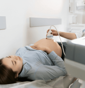
Ureteropelvic Junction Obstruction (UPJ Obstruction) is a blockage or narrowing at the junction between the renal pelvis and the ureter, preventing normal drainage of urine from the kidney to the bladder. The obstruction may be congenital, resulting from abnormal development during fetal growth, or acquired, due to scar tissue, stones, blood vessels, or previous surgeries. This condition can lead to progressive kidney damage if not treated and is commonly detected during childhood or prenatal ultrasound but may also present in adults.
Symptoms
Symptoms vary depending on the severity and whether the condition is unilateral or bilateral:
- Flank or abdominal pain (especially after fluid intake)
- Nausea and vomiting
- Hematuria (blood in the urine)
- Recurrent urinary tract infections
- Palpable abdominal mass in infants or children
- Failure to thrive (in infants)
- Hypertension
- Kidney stones
- Intermittent or chronic back pain
Causes
UPJ obstruction can be due to a number of congenital or acquired factors:
- Congenital causes (most common in children):
- Intrinsic narrowing of the UPJ
- Abnormal muscle development or insertion
- Presence of crossing blood vessels compressing the junction
- Acquired causes (more common in adults):
- Scar tissue from infections or surgeries
- Kidney stones obstructing the junction
- Trauma or inflammation
- Tumors compressing the urinary tract
Risk Factors
- Male gender (more commonly diagnosed in boys)
- Family history of urinary tract abnormalities
- Congenital urinary anomalies
- Previous kidney or ureter surgeries
- Recurrent urinary tract infections
- Kidney stones
- Hydronephrosis detected on prenatal ultrasound
Complications
- Hydronephrosis (swelling of the kidney due to urine retention)
- Progressive kidney damage or renal failure
- Kidney stones due to urinary stasis
- High blood pressure
- Recurrent urinary tract infections
- Pain and reduced quality of life
- Urinary leakage or rupture (in extreme untreated cases)
Prevention
UPJ obstruction cannot be prevented in congenital cases, but early detection and management can prevent complications:
- Prenatal ultrasounds can identify hydronephrosis before birth
- Timely evaluation of urinary tract symptoms in children and adults
- Regular follow-up imaging for at-risk individuals
- Prompt treatment of infections or stones to avoid secondary obstruction
- Stay hydrated to reduce risk of stone formation
Treatment Options in Korea
South Korea provides advanced and minimally invasive treatment options for UPJ Obstruction in both pediatric and adult patients:
- Observation: In mild, non-obstructive cases or infants with improving hydronephrosis
- Diuretic renal scan (MAG3 or DTPA): Used to assess kidney drainage and function
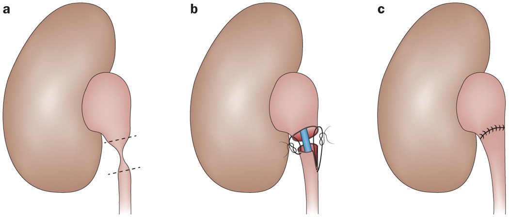
- Pyeloplasty: The most common surgical treatment, involving reconstruction of the UPJ to relieve obstruction
- Open pyeloplasty: Traditional surgical approach
- Laparoscopic or robotic-assisted pyeloplasty: Minimally invasive, with quicker recovery and excellent outcomes
- Endopyelotomy: Internal incision of the blockage using a ureteroscope or laser, often used in adults
- Stent placement: May be used temporarily to relieve obstruction
- Postoperative follow-up: Regular imaging to monitor kidney function and ensure long-term success
With high surgical precision, pediatric expertise, and comprehensive imaging services, Korean hospitals deliver world-class outcomes for UPJ obstruction treatment.