Overview
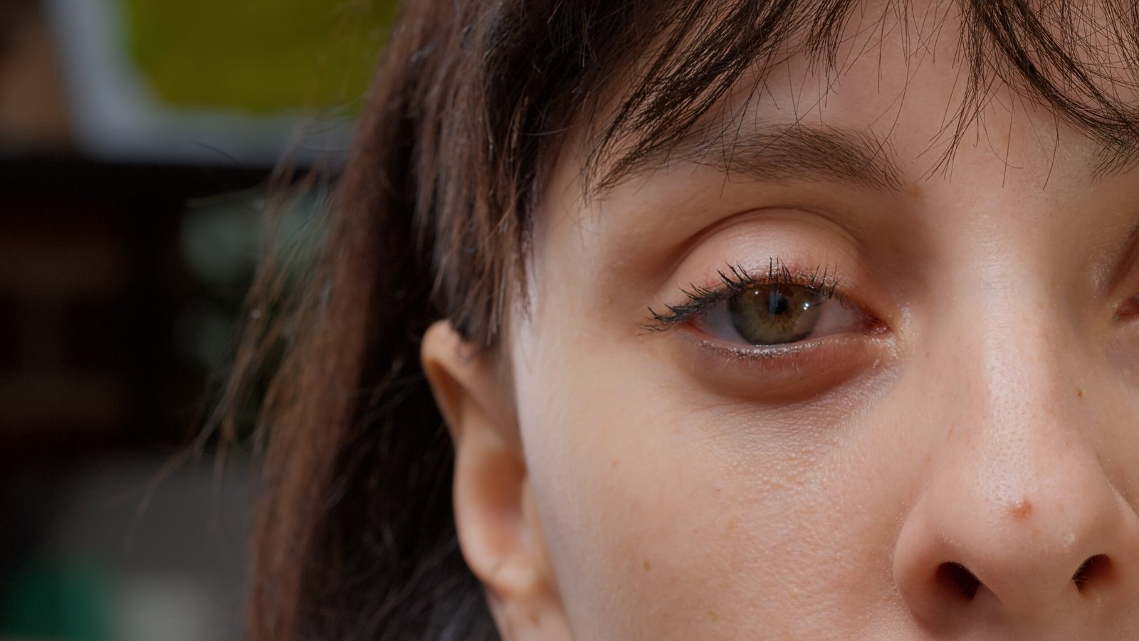
Synechia refers to an abnormal adhesion between parts of the eye, most commonly involving the iris sticking to either the lens (posterior synechia) or the cornea (anterior synechia). These adhesions can impair vision, increase intraocular pressure, and lead to complications such as glaucoma or cataracts. In Korea, ophthalmologists use advanced diagnostic and surgical techniques to manage synechia effectively, especially when detected early.
What is Synechia?
Synechia is a condition where tissue in the eye adheres abnormally to adjacent structures. There are two main types:
- Anterior Synechia: Adhesion between the iris and cornea.
- Posterior Synechia: Adhesion between the iris and the lens.
These adhesions may block normal fluid flow inside the eye, causing increased intraocular pressure and risking vision-threatening diseases.
Synechia is often a secondary condition, resulting from inflammation, trauma, surgery, or certain eye diseases.
Symptoms
Many cases are asymptomatic in the early stages but may eventually cause:
- Blurred or decreased vision
- Eye pain or discomfort
- Irregular pupil shape
- Redness or inflammation
- Sensitivity to light (photophobia)
- Increased intraocular pressure (if glaucoma develops)
Causes
- Uveitis or iritis (intraocular inflammation)
- Trauma or injury to the eye
- Eye surgery (especially cataract surgery)
- Infections (e.g., herpes simplex, tuberculosis)
- Congenital eye disorders
- Chemical or thermal burns
Risk Factors
- Autoimmune conditions (e.g., lupus, rheumatoid arthritis)
- Previous eye surgeries
- Eye infections or injuries
- Chronic uveitis
- Poorly controlled diabetes
- History of intraocular inflammation
Complications
If not treated early, synechia can lead to:
- Glaucoma (due to blocked drainage of aqueous humor)
- Cataracts
- Permanent vision loss
- Irregular pupil function
- Difficulty during future eye surgeries
Prevention
- Timely treatment of eye infections or inflammation
- Protective eyewear to prevent trauma
- Regular follow-up after eye surgeries
- Managing underlying autoimmune or systemic diseases
- Using prescribed anti-inflammatory eye drops as directed
Treatment Options in Korea
South Korea offers advanced eye care services and high success rates in managing synechia using both medical and surgical methods.
Diagnosis
- Slit-lamp examination
- Tonometry (to measure intraocular pressure)
- Ophthalmoscopy
- Anterior segment optical coherence tomography (AS-OCT)
- Ultrasound biomicroscopy
Medical Management
- Cycloplegic eye drops: To break adhesions and reduce inflammation
- Steroid eye drops: To control inflammation
- Intraocular pressure-lowering drops (if glaucoma is present)
- Antibiotics or antivirals (if infection is involved)
Surgical Treatment
- Synechiolysis: A procedure to break the adhesions, performed using a laser or microsurgical techniques
- Glaucoma surgery (e.g., trabeculectomy) if intraocular pressure remains high
- Cataract surgery: If synechia is associated with lens opacity
- Anterior segment reconstruction in complex cases
Leading Hospitals in Korea
- Seoul National University Hospital (SNUH) Eye Center
- Kim’s Eye Hospital, Seoul
- B&VIIT Eye Center
- Yonsei Severance Hospital Ophthalmology Department
Rehabilitation and Follow-up
- Regular eye exams post-treatment
- Monitoring for glaucoma
- Use of protective eyewear
- Patient education for managing underlying conditions