Overview
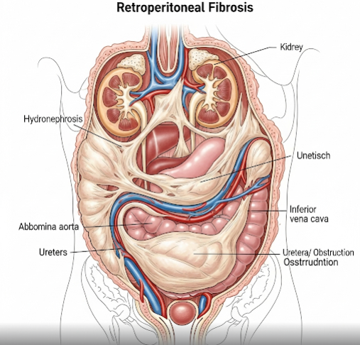
Retroperitoneal fibrosis (RPF) is a rare condition in which dense, fibrous tissue develops in the retroperitoneal space—the area behind the abdominal cavity where vital structures like the kidneys, ureters, and major blood vessels are located. This fibrous tissue can encase and obstruct the ureters, leading to kidney damage. In Korea, advanced diagnostic imaging and minimally invasive treatments make it possible to detect RPF early and manage it effectively, helping to preserve kidney function and overall health.
What is Retroperitoneal Fibrosis?
Retroperitoneal fibrosis occurs when abnormal fibrous tissue forms in the retroperitoneal space, often encasing nearby organs and structures. The exact cause is unknown in many cases (idiopathic RPF), but it can also result from infections, medications, autoimmune disorders, or cancer. If left untreated, the fibrosis can block urinary flow from the kidneys, leading to serious complications such as hydronephrosis and renal failure.
Symptoms
- Persistent abdominal or flank pain
- Back pain
- Lower extremity swelling
- Nausea and vomiting
- Weight loss
- Fatigue
- Decreased urine output (in advanced cases)
- Symptoms of kidney failure (if untreated)
Causes
- Idiopathic (unknown cause – most common)
- Autoimmune diseases (e.g., IgG4-related disease)
- Chronic infections (e.g., tuberculosis)
- Certain medications (e.g., ergot derivatives, beta-blockers)
- Abdominal or pelvic surgery history
- Cancers of the abdomen or pelvis
Risk Factors
- Middle-aged individuals (most cases occur between ages 40–60)
- Male gender (higher prevalence)
- Family history of autoimmune disease
- Long-term use of certain medications
- History of abdominal trauma or surgery
Complications
- Hydronephrosis (swelling of kidneys due to urine backup)
- Chronic kidney disease or renal failure
- Hypertension (due to kidney involvement)
- Lower limb edema
- Increased risk of infections
Prevention
- Regular monitoring if at risk due to medication use or autoimmune conditions
- Prompt treatment of infections and inflammatory conditions
- Avoidance of medications known to trigger fibrosis when possible
- Routine health check-ups with kidney function monitoring
Treatment Options in Korea
Korean hospitals use a multidisciplinary approach for RPF, involving nephrologists, urologists, and radiologists:
- Diagnostic Tools: CT scan, MRI, PET scan, and sometimes biopsy to confirm fibrosis and rule out cancer.
- Medical Therapy:
- Corticosteroids to reduce inflammation and slow fibrosis progression.
- Immunosuppressive drugs for autoimmune-related cases.
- Surgical Management:
- Ureterolysis (surgical freeing of the ureters from fibrous tissue) via minimally invasive laparoscopic or robotic techniques.
- Stent Placement: Ureteral stents or nephrostomy tubes to relieve urinary obstruction.
- Long-term Monitoring: Regular imaging and kidney function tests to ensure no recurrence or progression.
With Korea’s advanced imaging, robotic surgery capabilities, and tailored medical therapies, patients with retroperitoneal fibrosis can achieve significant symptom relief and maintain kidney function over the long term.