Overview
Intramuscular lipoma is a rare and distinctive type of benign soft tissue tumor composed of mature adipose (fat) cells that arise deep within skeletal muscle tissue. Unlike the more common superficial (subcutaneous) lipomas, which are typically well-circumscribed and easily movable beneath the skin, intramuscular lipomas grow within the muscle fibers themselves, often infiltrating between them. This infiltrative growth pattern makes intramuscular lipomas more challenging to diagnose and treat, as they can blend with surrounding muscle tissue and occasionally cause symptoms related to their size and location. Although these tumors are benign and do not metastasize, they may cause discomfort, functional impairment, or cosmetic concerns. In Korea, the management of intramuscular lipomas benefits from cutting-edge diagnostic imaging, histopathological expertise, and advanced surgical techniques that ensure effective treatment and functional preservation.
What is Intramuscular Lipoma?
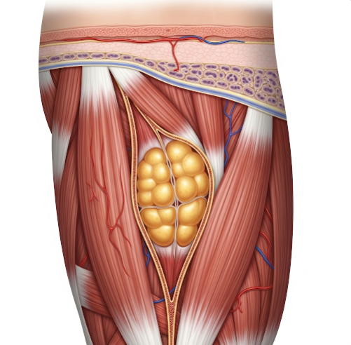
An intramuscular lipoma is a benign tumor consisting predominantly of mature adipocytes that originate within the muscle belly. Unlike conventional lipomas that are encapsulated and located just beneath the skin, intramuscular lipomas exhibit an infiltrative growth pattern, interdigitating between muscle fibers and sometimes extending along fascial planes. This growth pattern may cause the lesion to appear ill-defined on clinical examination and imaging.
Intramuscular lipomas most frequently involve large muscle groups such as the thigh (quadriceps), shoulder muscles (deltoid), back muscles (latissimus dorsi), and the forearm, but they can occur in any skeletal muscle. These tumors tend to grow slowly over months or years, and their size can vary from small nodules to large masses weighing several kilograms.
Symptoms
Many intramuscular lipomas are initially asymptomatic and detected incidentally during imaging for unrelated reasons. Symptoms generally arise as the tumor enlarges or exerts pressure on adjacent structures, including nerves, blood vessels, or muscle tissue itself. Common symptoms include:
- Palpable deep-seated mass: Often a firm, painless lump felt within a muscle. Because of its deep location, it may not be easily noticeable externally.
- Localized pain or discomfort: May result from nerve compression or irritation within the muscle.
- Muscle weakness or cramping: When the tumor disrupts normal muscle contraction or compresses nerves controlling the muscle.
- Restricted range of motion: Particularly if the lipoma is near a joint or involves muscles important for limb movement.
- Swelling or a sense of fullness: In the affected area, sometimes noticeable under the skin.
- Rare neurological symptoms: Such as numbness or tingling if peripheral nerves are compressed.
Causes
The precise cause of intramuscular lipomas remains unclear. Several hypotheses and contributing factors have been proposed:
- Genetic predisposition: Some studies suggest a hereditary tendency to develop lipomas, though no specific gene mutations have been conclusively identified.
- Local trauma or muscle injury: Repeated microtrauma or a single significant injury might trigger adipocyte proliferation and tumor formation in muscle tissue.
- Metabolic factors: Abnormalities in fat metabolism or adipocyte regulation could contribute.
- Age-related changes: Most intramuscular lipomas occur in middle-aged adults, possibly due to cumulative environmental or metabolic effects.
Risk Factors
Several factors may increase the risk of developing intramuscular lipomas or contribute to their growth:
- Age: Most commonly diagnosed in individuals aged 40 to 60 years.
- History of trauma: Previous muscle injury or repeated mechanical stress.
- Obesity: Excessive body fat might predispose to abnormal fat tissue growth.
- Family history: Genetic predisposition or familial lipomatosis syndromes.
- Other soft tissue tumors: Presence of multiple lipomas or other benign tumors.
Complications
Although intramuscular lipomas are benign and do not metastasize, they can cause complications if untreated or large:
- Functional impairment: As the lipoma infiltrates muscle fibers, it may reduce muscle strength and impair normal movement.
- Chronic pain: From compression of nerves or irritation of muscle tissue.
- Compression of adjacent structures: Large tumors can press on blood vessels or nerves causing swelling, numbness, or weakness.
- Recurrence after surgery: Due to the infiltrative nature, incomplete removal may lead to regrowth.
- Diagnostic confusion: They can mimic malignant soft tissue tumors (e.g., well-differentiated liposarcomas), necessitating careful evaluation.
Prevention
There are no specific measures to prevent intramuscular lipomas due to the unclear etiology. However, general recommendations to promote muscle and tissue health include:
- Avoiding repetitive muscle trauma: Using protective measures during activities that stress muscles.
- Maintaining healthy body weight: To potentially reduce abnormal fat accumulation.
- Early medical evaluation: Prompt assessment of any unusual muscle lumps or persistent discomfort to enable early diagnosis.
Treatment Options in Korea
Korea’s healthcare system offers advanced, multidisciplinary approaches to diagnosing and treating intramuscular lipomas:
- Diagnostic Imaging:
- Magnetic Resonance Imaging (MRI): The most sensitive modality, MRI provides detailed visualization of the lipoma’s size, exact location, and infiltration into muscle fibers. Intramuscular lipomas typically appear as well-defined or infiltrative lesions with fat signal intensity.
- Ultrasound: Useful for initial assessment, distinguishing cystic versus solid masses, and guiding biopsies.
- Computed Tomography (CT): Occasionally used for detailed anatomical evaluation, especially in complex cases.
- Histopathological Examination:
- Core needle biopsy: Performed when imaging cannot definitively exclude malignancy, providing tissue for microscopic examination.
- Surgical specimen analysis: Confirmatory diagnosis is achieved after excision.
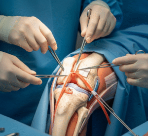
- Surgical Management:
- Complete surgical excision is the treatment of choice. Due to the infiltrative growth, surgeons aim to remove the lipoma along with some surrounding muscle tissue to minimize recurrence risk.
- Minimally invasive surgical techniques are employed where possible to reduce tissue damage and preserve muscle function.
- Korean orthopedic and plastic surgeons are skilled in preserving muscle integrity while achieving clear margins.
- Postoperative Care and Rehabilitation:
- Physical therapy: Customized rehabilitation programs to restore muscle strength, flexibility, and function.
- Regular follow-up: Imaging surveillance to detect potential recurrence.
- Pain management: If residual discomfort occurs post-surgery.
- Multidisciplinary Team Approach:
- Collaboration between orthopedic surgeons, radiologists, pathologists, and rehabilitation specialists ensures accurate diagnosis, optimal surgical planning, and comprehensive postoperative care.