Overview
Branch Retinal Vein Occlusion (BRVO) is an eye condition in which one of the smaller branches of the retinal veins becomes blocked, leading to impaired blood drainage from the retina. This blockage can cause retinal swelling, bleeding, and vision disturbances, and is a significant cause of sudden, painless vision loss in adults.
In Korea, BRVO is managed by ophthalmologists and retinal specialists using advanced diagnostic imaging, laser therapy, anti-VEGF injections, and comprehensive eye care. Korean eye hospitals are equipped with cutting-edge retina imaging technology to monitor disease progression and optimize treatment outcomes.
What is Branch Retinal Vein Occlusion (BRVO)?
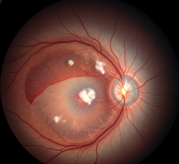
BRVO occurs when a branch of the central retinal vein becomes obstructed, usually at points where arteries and veins cross. The blockage causes retinal blood vessels to leak fluid, leading to macular edema, hemorrhages, and sometimes vision impairment.
BRVO is classified based on the location of the blockage:
- Superotemporal BRVO: Upper outer retina
- Inferotemporal BRVO: Lower outer retina
- Other branch locations depending on retinal anatomy
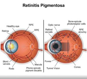
Unlike central retinal vein occlusion (CRVO), BRVO affects only a portion of the retina, which usually results in localized vision changes rather than complete loss of vision.
Symptoms
Symptoms of BRVO may develop suddenly or gradually, depending on severity:
- Blurred or distorted vision in part of the visual field
- Dark spots or floaters in vision
- Sudden loss of central vision in severe cases
- Visual field defects, often in the upper or lower quadrant
- Usually painless, but sudden changes should be evaluated promptly
Some patients may not notice early symptoms, making routine eye examinations crucial, especially for individuals with risk factors.
Causes
BRVO occurs primarily due to obstruction of a retinal vein, often at arteriovenous crossing points. Contributing factors include:
- Hypertension (high blood pressure): Increases risk of vascular damage
- Atherosclerosis: Hardening or thickening of retinal arteries compressing veins
- Diabetes mellitus: Damages blood vessels, leading to occlusion
- Glaucoma: Elevated intraocular pressure may affect retinal circulation
- Hyperlipidemia: High cholesterol contributes to vascular blockage
- Blood clotting disorders: Increases risk of venous occlusion
Risk Factors
- Age: Most common in individuals over 50 years
- Hypertension and cardiovascular disease
- Diabetes mellitus
- Hyperlipidemia
- Smoking
- Obesity and sedentary lifestyle
- Family history of vascular or retinal disorders
Complications
BRVO can lead to several vision-threatening complications if untreated:
- Macular edema: Swelling in the central retina causing vision loss
- Neovascularization: Abnormal blood vessel growth that may cause bleeding or glaucoma
- Retinal hemorrhages
- Permanent vision loss in severe or chronic cases
- Secondary glaucoma due to new vessel formation
Prevention
While some risk factors cannot be changed, BRVO risk can be reduced with lifestyle and medical interventions:
- Blood pressure control through diet, exercise, and medications
- Blood sugar management for diabetic patients
- Cholesterol and lipid regulation
- Avoiding smoking and alcohol abuse
- Regular eye check-ups for early detection
- Maintaining a healthy weight and active lifestyle
Treatment Options in Korea
Diagnosis
Early detection is essential for preserving vision:
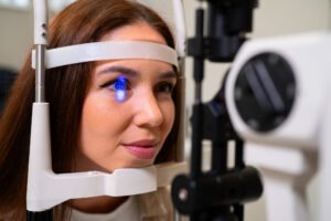
- Fundus examination: Direct visualization of retinal vessels and hemorrhages
- Optical Coherence Tomography (OCT): Measures retinal thickness and detects macular edema
- Fluorescein angiography: Visualizes blood flow and identifies blocked veins
- Visual acuity and field testing to monitor functional impact
Medical Management
- Observation: Mild cases without macular edema may be monitored
- Anti-VEGF injections: Medications such as ranibizumab, bevacizumab, or aflibercept reduce macular edema and improve vision
- Corticosteroid injections: Reduce inflammation and retinal swelling
- Management of underlying conditions: Hypertension, diabetes, and hyperlipidemia must be controlled
Laser Therapy
- Focal laser photocoagulation: Seals leaking blood vessels to prevent further edema
- Scatter (panretinal) laser therapy: Used in cases with neovascularization to prevent complications
Surgical Options
Surgery is rare but may be considered for persistent macular edema or complications:
- Vitrectomy: Removes vitreous hemorrhage and relieves traction
- Surgical interventions for glaucoma secondary to BRVO
Supportive Care
- Routine follow-up to monitor retinal changes
- Vision rehabilitation if vision loss persists
- Patient education on recognizing sudden changes or bleeding
Prognosis
The prognosis of BRVO depends on severity, treatment, and underlying health conditions:
- Mild cases often retain good vision with careful monitoring
- Macular edema responds well to anti-VEGF therapy in most patients
- Early detection and treatment in Korea with advanced retinal care significantly improve visual outcomes
- Long-term follow-up ensures early intervention for recurrence or neovascular complications
Korean eye hospitals provide state-of-the-art diagnostic tools, intravitreal therapies, laser treatments, and comprehensive retinal care, offering patients with BRVO the best chance for preserving vision and preventing complications.