Overview
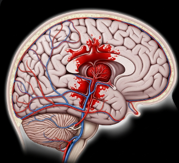
A brain hemorrhage, also called intracerebral hemorrhage, occurs when a blood vessel in the brain ruptures, causing bleeding into surrounding brain tissue. This bleeding increases intracranial pressure and can severely damage brain cells, potentially leading to stroke, neurological deficits, or death.
In Korea, brain hemorrhage is treated in neurosurgery, neurology, and intensive care units using advanced imaging, emergency interventions, and surgical techniques. Korean hospitals focus on rapid diagnosis, controlling bleeding, managing complications, and providing rehabilitation, aiming to minimize long-term neurological damage.
What is a Brain Hemorrhage?
A brain hemorrhage is a type of stroke caused by bleeding rather than a blockage of blood flow. It can be classified based on location:
- Intracerebral hemorrhage (ICH): Bleeding within brain tissue itself
- Subarachnoid hemorrhage (SAH): Bleeding into the space between the brain and skull, often from a ruptured aneurysm
- Subdural hemorrhage: Bleeding between the dura mater and brain surface
- Epidural hemorrhage: Bleeding between the skull and dura mater, often trauma-related
Brain hemorrhages can cause sudden neurological deficits, and the severity depends on bleed size, location, and speed of onset.
Symptoms
Symptoms may develop suddenly or progress gradually, depending on hemorrhage type and size:
- Sudden, severe headache, often described as the worst ever experienced
- Nausea and vomiting
- Weakness or numbness on one side of the body
- Difficulty speaking or understanding speech (aphasia)
- Vision problems, including double vision or loss of vision
- Loss of balance or coordination
- Seizures
- Altered consciousness, ranging from confusion to coma
In cases of subarachnoid hemorrhage, patients may experience a “thunderclap” headache, stiff neck, and sensitivity to light.
Causes
Brain hemorrhages occur due to rupture of weakened blood vessels, which may result from:
- High blood pressure (hypertension): Leading cause of intracerebral hemorrhage
- Aneurysm rupture: Weak spots in arteries that balloon and burst
- Arteriovenous malformations (AVMs): Abnormal tangles of blood vessels
- Head trauma: Accidents or falls causing epidural or subdural hemorrhage
- Blood clotting disorders: Hemophilia or anticoagulant medications
- Liver disease or severe infections affecting coagulation
- Brain tumors: Malignant tumors can bleed spontaneously
Risk Factors
- Hypertension: Chronic high blood pressure increases vessel fragility
- Age: Risk increases in older adults
- Smoking and alcohol consumption
- Use of anticoagulant or antiplatelet medications
- Family history of stroke or brain aneurysms
- Trauma or high-impact accidents
- Chronic medical conditions: Diabetes, kidney disease, or vascular disorders
Complications
Brain hemorrhages can lead to severe, life-threatening complications:
- Increased intracranial pressure, compressing brain tissue
- Brain herniation, a life-threatening displacement of brain tissue
- Permanent neurological deficits, including paralysis, speech difficulties, or cognitive impairment
- Seizures and ongoing epilepsy
- Hydrocephalus, due to obstruction of cerebrospinal fluid
- Coma or death in severe cases
Prevention
Preventive measures focus on reducing risk factors and managing underlying conditions:
- Blood pressure control: Medications, dietary changes, and exercise
- Avoiding tobacco and excessive alcohol
- Monitoring and managing chronic diseases such as diabetes or kidney disorders
- Safe use of anticoagulants with regular medical supervision
- Protective measures to prevent head trauma
- Early detection and treatment of aneurysms or AVMs
Treatment Options in Korea
Diagnosis
Rapid and accurate diagnosis is critical:
- CT scan (computed tomography): Quick identification of bleeding location and size
- MRI (magnetic resonance imaging): Detailed imaging of brain tissue
- CT or MR angiography: Identifies aneurysms or vascular malformations
- Blood tests: Assess coagulation status and underlying conditions
- Neurological assessment: Evaluates consciousness, motor function, and cognitive status
Medical Management
- Blood pressure management: Reduce further bleeding risk
- Medications:
- Anticonvulsants to prevent seizures
- Osmotic agents like mannitol to reduce intracranial pressure
- Pain relief and supportive therapy
- Monitoring in ICU: Continuous neurological observation and vital support
Surgical Management
Surgery may be necessary in certain cases:
- Craniotomy: Removing clot or hematoma to relieve pressure
- Endovascular coiling or clipping: For aneurysm-related hemorrhage
- Decompressive craniectomy: Relieves severe swelling and prevents brain herniation
- AVM resection: Removal of abnormal blood vessels to prevent rebleeding
Supportive Care
- Rehabilitation: Physical, occupational, and speech therapy to recover function
- Psychological support: Counseling for patients and families
- Nutritional and fluid management
- Long-term monitoring through imaging to detect recurrence or complications
Prognosis
The prognosis depends on bleed size, location, patient age, and timeliness of treatment:
- Small, promptly treated hemorrhages may have good outcomes with minimal deficits
- Large or untreated hemorrhages carry a high risk of permanent neurological damage or death
- Korean hospitals provide state-of-the-art neurosurgical, intensive care, and rehabilitation services, improving survival rates and functional recovery
- Early intervention with surgical or medical management significantly reduces complications and enhances quality of life
With advanced diagnostic tools, emergency care, and multidisciplinary treatment teams, Korea offers world-class care for brain hemorrhage, aiming to save lives, minimize disability, and restore patient independence.