Overview
Breast screening is a preventive medical procedure that uses mammography (X-ray imaging) to detect breast cancer at an early stage, often before symptoms develop. Early detection is critical to improve treatment outcomes and survival rates.
Importance of breast screening:
- Detects breast cancer before it becomes palpable.
- Reduces mortality by identifying tumors early.
- Guides treatment planning and intervention.
- Enables monitoring of high-risk individuals for timely detection.
In South Korea, mammography is widely available in hospitals, breast centers, and imaging clinics, supported by national screening programs for women in specific age groups. Modern digital mammography and tomosynthesis improve accuracy and early detection.
Why It’s Done
Mammograms are performed primarily to detect breast cancer early and monitor breast health.
Common indications include:
- ➤ Routine screening: Recommended for women aged 40–69 years, with intervals depending on national guidelines.
- ➤ High-risk individuals: Family history of breast cancer, BRCA1/2 gene mutations, or prior chest radiation.
- ➤ Evaluation of breast symptoms: Lumps, pain, nipple discharge, or skin changes.
- ➤ Follow-up after breast cancer treatment to monitor recurrence.
Benefits for patients:
- ✔️ Early detection leads to less aggressive treatment and better survival.
- ✔️ Identifies tumors too small to feel, often before metastasis.
- ✔️ Provides baseline imaging for future comparisons.
Alternatives
While mammography is the gold standard for screening, other options exist depending on patient age, risk, or breast density:
- ➤ Breast ultrasound: Useful for dense breast tissue or to evaluate specific lumps.
- ➤ MRI (Magnetic Resonance Imaging): Recommended for high-risk patients; more sensitive but costly.
- ➤ Clinical breast examination: Performed by healthcare professionals to detect abnormalities.
- ➤ Self-examination: Increases awareness but cannot replace imaging.
Key point: Mammography remains the most effective method for population-wide screening, while alternatives are complementary for high-risk or special cases.
Preparation
Preparation for a mammogram is simple but important for optimal results.
Steps include:
- ✅ Schedule timing: Avoid screening during menstrual period when breasts may be tender.
- ✅ Avoid deodorants or powders: These can appear as artifacts on the X-ray.
- ✅ Wear comfortable clothing: A two-piece outfit is convenient for upper body exposure.
- ✅ Provide medical history: Include prior surgeries, implants, family history, or previous imaging.
- ✅ Previous mammogram images: Bring past mammograms for comparison if available.
Important: Proper preparation ensures clear, accurate imaging and reduces the need for repeat scans.
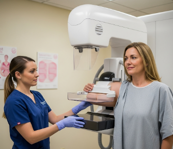
How It’s Done
Mammography is a non-invasive imaging procedure performed by trained radiology technicians.
Step-by-step process:
- Patient positioning: One breast at a time is placed on the mammography unit.
- Compression: Breast is gently compressed to spread tissue for clear imaging.
- X-ray exposure: Images captured from different angles – usually top-to-bottom and side-to-side.
- Additional views: Taken if any area requires closer evaluation.
- Image review: Radiologist examines images for masses, calcifications, or asymmetries.
Duration: Approximately 15–30 minutes for the entire procedure.
Comfort: Compression may cause temporary discomfort but is necessary for image clarity.
Recovery & Follow-up
Mammograms do not require recovery; patients can resume normal activities immediately.
Post-procedure:
- Mild breast tenderness or redness may occur but resolves quickly.
- No restrictions on activities.
- Results usually available within a few days to a week.
Follow-up:
- Normal results: Continue routine screening as per age and risk guidelines.
- Abnormal findings: Additional imaging (ultrasound, MRI) or biopsy may be recommended.
- High-risk patients: More frequent surveillance or preventive measures may be advised.
Important: Maintaining a record of mammogram results helps track changes over time and facilitates early intervention.
Possible Complications
Mammography is very safe, with minimal risks:
- ⚠️ Radiation exposure: Extremely low; modern digital mammography uses minimal X-ray doses.
- ⚠️ Discomfort or pain: Temporary, during breast compression.
- ⚠️ False positives: May require additional tests, causing anxiety.
- ⚠️ False negatives: Rare, especially in dense breast tissue; cancer may be missed.
- ⚠️ Skin irritation or bruising: Rare, usually mild.
In South Korea, quality assurance programs and advanced imaging technologies minimize risks and improve accuracy.
Treatment Options / Clinical Relevance in Korea
Breast screening in South Korea is part of national cancer prevention programs.
Key features:
- 🏥 Government-supported screening for women aged 40–69, often free or subsidized.
- 🏥 Digital mammography and tomosynthesis for improved detection, especially in dense breasts.
- 🏥 Integrated breast centers provide imaging, biopsy, and oncology consultations.
- 🏥 Follow-up programs ensure high-risk patients receive timely care.
- 🏥 Educational campaigns raise awareness about early detection and preventive measures.
Hospitals and centers offering breast screening:
- Asan Medical Center – Comprehensive breast imaging and oncology services
- Samsung Medical Center – Advanced mammography and 3D breast tomosynthesis
- Seoul National University Hospital – National breast cancer screening program
- Local public health centers – Accessible mammography screening
Highlights in Korea:
- ✔️ Early detection improves survival rates and reduces aggressive treatment.
- ✔️ National programs ensure wide accessibility and high-quality care.
- ✔️ Integration with digital records allows long-term tracking of patient breast health.
- ✔️ Advanced imaging enhances accuracy in women with dense breast tissue.
Highlights
- ➤ Mammography is the gold standard for breast cancer screening, enabling early detection.
- ➤ Recommended for women aged 40–69 or high-risk individuals.
- ➤ Preparation is simple: avoid deodorants, wear comfortable clothing, and provide prior imaging.
- ➤ Procedure involves breast compression and X-ray imaging from multiple angles.
- ➤ Recovery is immediate, with results reviewed by radiologists.
- ➤ Risks are minimal, including mild discomfort or rare false positives/negatives.
- ➤ South Korea provides comprehensive, government-supported breast screening programs, with advanced digital imaging and integrated follow-up care.