Overview
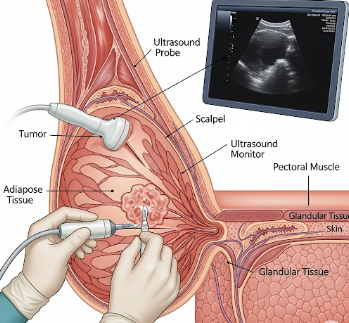
Imaging-Assisted Wide Local Excision (WLE) is a surgical technique used to remove tumors with a margin of healthy tissue while preserving as much normal tissue as possible. The “imaging-assisted” component uses real-time imaging guidance (ultrasound, MRI, or mammography) to precisely locate the tumor and plan excision.
South Korea is recognized for advanced surgical oncology, high-resolution imaging, and minimally invasive tumor removal techniques, making it a preferred destination for patients seeking safe, accurate, and cosmetically optimized WLE procedures.
What is Imaging-Assisted Wide Local Excision?
Imaging-assisted WLE combines:
- Precise tumor localization using imaging modalities
- Surgical removal of tumor with safe margins
- Preservation of surrounding healthy tissue for cosmetic and functional outcomes
- Intraoperative imaging to confirm complete excision
Indications include:
- Early-stage breast cancer or other soft tissue tumors
- Benign but suspicious lesions requiring complete excision
- Tumors in cosmetically sensitive areas where tissue preservation is important
- Patients requiring accurate excision without reoperation
What are the Benefits?
- Accurate tumor removal → Minimizes residual tumor risk
- Preserves healthy tissue → Better cosmetic and functional outcomes
- Minimally invasive approach → Smaller incision, less scarring
- Real-time imaging guidance → Reduces the need for re-excision
- Improved surgical planning → Enables optimal incision placement and tissue conservation
- Expert surgical care in Korea → High precision, safety, and low complication rates
Procedure Details
1) How should I prepare for Imaging-Assisted WLE?
- Preoperative evaluation → Imaging studies (ultrasound, MRI, or mammography) to map tumor location
- Medical history review → Assess comorbidities, medications, allergies
- Medication guidance → Blood thinners may need temporary adjustment
- Fasting → 6–8 hours prior to surgery if general anesthesia is planned
- Pre-procedure consultation → Discuss procedure steps, expected outcomes, and cosmetic considerations
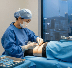
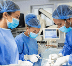
2) What happens during the procedure?
- Anesthesia → Local anesthesia with sedation or general anesthesia depending on tumor size and location
- Patient positioning → Optimized for tumor access and imaging
- Imaging guidance → Real-time imaging used to locate tumor accurately
- Surgical steps →
- Small incision made over the tumor site
- Tumor excised with a margin of healthy tissue
- Intraoperative imaging confirms complete removal
- Tissue may be sent for frozen section pathology if necessary
- Incision closed with minimal cosmetic disruption
- Duration → Typically 60–120 minutes depending on tumor size and complexity
- Monitoring → Continuous monitoring of vital signs and surgical field
3) What happens after Imaging-Assisted WLE?
- Immediate recovery → Observation for a few hours to overnight depending on anesthesia and procedure complexity
- Pain management → Mild analgesics prescribed for post-operative discomfort
- Activity restrictions → Avoid heavy lifting and strenuous activity for 1–2 weeks
- Wound care → Keep incision clean and dry; follow-up dressing changes as advised
- Follow-up visits → Pathology review, incision check, and planning for adjuvant therapy if needed
Risks / Benefits
Risks
- ➤ Infection at the surgical site
- ➤ Bleeding or hematoma formation
- ➤ Pain or swelling at the incision site
- ➤ Need for re-excision if margins are not clear
- ➤ Rare injury to surrounding structures depending on tumor location
- ➤ Temporary cosmetic asymmetry or scarring
Benefits
- ➤ Accurate removal of tumor with clear margins
- ➤ Preservation of healthy tissue and cosmetic outcome
- ➤ Reduced risk of reoperation due to imaging guidance
- ➤ Minimally invasive with shorter recovery
- ➤ Expert surgical care in Korea ensures optimal precision and safety
Recovery and Outlook
- Immediate recovery → Mild soreness and swelling around incision
- Short-term follow-up → Usually within 1–2 weeks to assess wound healing and pathology results
- Return to normal activity → Light activity after a few days; full recovery in 2–4 weeks
- Long-term outlook → High likelihood of complete tumor removal with good cosmetic results
- Post-procedure care → Regular imaging and clinical follow-up to monitor for recurrence
- Integration with oncology care → Adjuvant therapy such as radiation or chemotherapy may be planned based on pathology
South Korea provides comprehensive follow-up care, imaging services, and cosmetic surgery support to ensure optimal recovery and patient satisfaction.
When To Call the Doctor
Contact your surgeon immediately if you notice:
- ⚠️ Fever, chills, or signs of infection
- ⚠️ Excessive bleeding or discharge from the incision
- ⚠️ Severe or worsening pain not relieved by medications
- ⚠️ Skin changes, redness, or swelling around the surgical site
- ⚠️ Any concern regarding cosmetic appearance or wound healing
Best Korea Option / Process
South Korea is a leading destination for Imaging-Assisted WLE due to:
- Expert surgical oncologists and breast/soft tissue surgeons
- High-resolution imaging technology for intraoperative guidance
- Minimally invasive techniques with optimal cosmetic outcomes
- Comprehensive post-operative care and follow-up
- International patient services → Consultation, language support, and treatment coordination
Top Hospitals for Imaging-Assisted WLE in Korea:
- Asan Medical Center, Seoul – Advanced intraoperative imaging and tumor excision
- Samsung Medical Center – Precision-guided excisions with frozen section pathology
- Seoul National University Hospital (SNUH) – Integrated oncologic and surgical care
- Yonsei Severance Hospital – Multidisciplinary tumor management with cosmetic expertise
👉 For patients requiring precise tumor removal with optimal cosmetic and functional outcomes, Imaging-Assisted Wide Local Excision in Korea offers a safe, highly accurate, and expert surgical solution.