Overview
Cavernous hemangioma, also called cavernous malformation or cavernoma, is a cluster of abnormal, dilated blood vessels that can occur in the brain, spinal cord, or other organs such as the liver and eyes. These lesions may remain asymptomatic for years, but they can also cause neurological symptoms, seizures, or hemorrhage if located in the central nervous system. In Korea, diagnosis and treatment of cavernous hemangiomas are advanced, with specialized neuroimaging, minimally invasive neurosurgery, and radiosurgery options available.
What is Cavernous Hemangioma?
A cavernous hemangioma is a benign vascular malformation made of enlarged capillary-like vessels filled with blood. Unlike cancer, it does not spread, but its location can cause significant health problems. When found in the brain or spinal cord, cavernous hemangiomas can leak or bleed, leading to neurological symptoms. They can also occur in the liver, where they are usually harmless but sometimes require monitoring.
Symptoms
- Brain/Spinal Cavernous Hemangioma:
- Seizures
- Headaches
- Weakness or numbness in arms/legs
- Vision or speech disturbances
- Balance or coordination problems
- Sudden neurological deficits due to hemorrhage
- Liver Cavernous Hemangioma:
- Often asymptomatic
- Abdominal pain or fullness (in large lesions)
- Rarely bleeding into the abdominal cavity
Causes
- Genetic mutations: Some cases are hereditary, caused by mutations in genes such as CCM1, CCM2, or CCM3.
- Sporadic cases: Many occur without family history.
- Radiation exposure: Rarely, radiation therapy may induce vascular malformations.
Risk Factors
- Family history of cavernous malformations
- Previous brain radiation
- Certain genetic syndromes
- Female sex (liver hemangiomas are more common in women)
Complications
- Intracerebral hemorrhage (bleeding in the brain)
- Stroke-like symptoms
- Recurrent seizures
- Permanent neurological deficits (depending on bleed location)
- Rare liver rupture with internal bleeding (for hepatic cavernous hemangioma)
Prevention
- No guaranteed prevention, but people with family history can undergo genetic counseling and MRI screening.
- Early monitoring of symptoms such as seizures or unexplained headaches.
- Avoiding unnecessary head trauma.
Treatment Options in Korea
Korea provides advanced diagnosis and treatment options for cavernous hemangiomas, particularly in brain and spinal cases.
1. Diagnosis
- MRI with contrast is the gold standard, especially for brain/spinal lesions.
- CT scan may detect hemorrhage.
- Abdominal ultrasound or CT for liver cavernous hemangiomas.
2. Observation & Monitoring
- If asymptomatic and small, doctors may recommend regular MRI follow-ups.
3. Medications
- Anti-seizure drugs for patients with epilepsy caused by cavernous malformations.
- Pain management for headaches.
4. Surgery
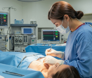
- Microsurgical resection is recommended if the lesion is causing repeated bleeding, seizures, or progressive neurological symptoms.
- Advanced neuronavigation and minimally invasive neurosurgical techniques are widely available in Korean hospitals.
5. Radiosurgery
- Gamma Knife Radiosurgery (GKS) is available in Korea for deep or inoperable cavernous hemangiomas, especially in the brainstem.
6. Liver Hemangioma Treatment
- Usually, no treatment is needed unless the lesion is very large or symptomatic.
- Surgery (resection) or embolization may be considered in rare complicated cases.