Overview
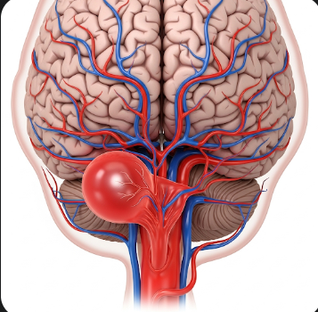
A brain aneurysm, also called a cerebral aneurysm, is a weak or thin spot in the wall of a brain artery that bulges or balloons out, forming a sac-like structure. While many brain aneurysms remain small and asymptomatic, rupture can lead to life-threatening hemorrhagic stroke or subarachnoid hemorrhage.
In Korea, brain aneurysms are treated in neurosurgery and interventional neuroradiology centers using cutting-edge imaging, minimally invasive endovascular techniques, and advanced surgical methods. The focus is on early detection, prevention of rupture, and prompt intervention to minimize complications and improve patient outcomes.
What is a Brain Aneurysm?
A brain aneurysm develops when the wall of an artery in the brain weakens, allowing blood to push outward, forming a balloon-like bulge. Aneurysms vary in size:
- Small (<7 mm) – often asymptomatic
- Medium (7–12 mm) – may cause symptoms depending on location
- Large (13–24 mm) – higher risk of rupture
- Giant (≥25 mm) – can compress surrounding brain tissue and nerves
Unruptured aneurysms may be discovered incidentally during imaging for unrelated issues. Ruptured aneurysms, however, cause sudden, severe neurological symptoms and require immediate medical attention.
Symptoms
Unruptured brain aneurysms often do not produce symptoms, but when symptoms occur, they may include:
- Headaches or localized pain above or behind the eye
- Visual disturbances, such as double vision or drooping eyelids
- Numbness or weakness on one side of the face or body
- Difficulty speaking or cognitive changes
Ruptured brain aneurysms present dramatically:
- Sudden, severe headache (“thunderclap headache”)
- Nausea and vomiting
- Stiff neck
- Sensitivity to light (photophobia)
- Loss of consciousness or confusion
- Seizures
- Neurological deficits depending on bleeding location
Causes
Brain aneurysms are primarily caused by structural weakness in blood vessel walls, and multiple factors contribute:
- Congenital defects: Weak arterial walls present from birth
- High blood pressure (hypertension): Increased stress on vessel walls
- Atherosclerosis: Plaque buildup weakening arteries
- Trauma or injury: Head injuries that damage vessels
- Infections: Rarely, arterial infections may contribute
- Vascular inflammation: Conditions like vasculitis
- Genetic disorders: Polycystic kidney disease or connective tissue disorders
Risk Factors
- Age: Most common in adults over 40
- Gender: Slightly higher prevalence in women
- Family history: Increased risk if first-degree relatives have aneurysms
- Hypertension: Chronic high blood pressure
- Smoking: Strongly associated with aneurysm formation and rupture
- Alcohol or drug use: Especially cocaine, which increases blood pressure acutely
- Previous aneurysm or cerebrovascular disease
Complications
Complications from brain aneurysms are severe and potentially life-threatening:
- Rupture leading to subarachnoid hemorrhage
- Stroke and brain damage due to bleeding
- Hydrocephalus from blood accumulation in brain ventricles
- Vasospasm: Narrowing of nearby arteries causing secondary stroke
- Permanent neurological deficits such as paralysis, speech difficulties, or cognitive impairment
- Death if untreated or if rupture is massive
Prevention
Preventive measures aim to reduce risk factors and detect aneurysms early:
- Blood pressure management: Regular monitoring and medication adherence
- Smoking cessation to reduce arterial stress
- Healthy lifestyle: Balanced diet, regular exercise, and weight control
- Avoiding illicit drugs that increase vascular strain
- Screening for high-risk individuals: Family history or genetic predisposition
- Early evaluation of neurological symptoms such as sudden severe headache, vision changes, or numbness
Treatment Options in Korea
Diagnosis
Diagnosis requires advanced imaging to identify size, location, and rupture risk:
- CT scan (computed tomography): Detects bleeding from ruptured aneurysms
- MRI (magnetic resonance imaging): Identifies unruptured aneurysms
- CT or MR angiography: Detailed images of blood vessels
- Digital subtraction angiography (DSA): Gold standard for evaluating aneurysm anatomy
Medical Management
- Observation and monitoring: Small, unruptured aneurysms may be monitored with regular imaging
- Blood pressure control: Reduces risk of rupture
- Lifestyle modifications: Smoking cessation, diet, and exercise
- Medications: Pain relief and prevention of vasospasm in ruptured cases
Surgical Management
Korea offers advanced surgical and minimally invasive options:
- Microsurgical clipping: Open surgery to place a clip at the aneurysm base
- Endovascular coiling: Minimally invasive procedure inserting coils to promote clotting and prevent rupture
- Flow diversion: Stents redirect blood flow away from aneurysm for large or complex cases
- Bypass surgery: Rarely used for complex vascular anatomy
Supportive Care
- ICU monitoring for ruptured aneurysms
- Rehabilitation: Physical, occupational, and speech therapy for neurological deficits
- Psychological support: Counseling for anxiety or post-stroke depression
- Regular follow-up imaging to monitor for recurrence or new aneurysms
Prognosis
The prognosis depends on size, location, rupture status, and timeliness of treatment:
- Unruptured aneurysms: High survival and excellent outcomes with early intervention
- Ruptured aneurysms: Mortality rates can be high without immediate treatment; early surgical or endovascular intervention significantly improves survival
- Neurological recovery: Rehabilitation in Korea is highly advanced, optimizing function after stroke or brain injury
- Long-term management: Blood pressure control, lifestyle modifications, and periodic imaging reduce recurrence risk
Korean hospitals provide state-of-the-art neurosurgery, interventional radiology, and rehabilitation services, ensuring patients with brain aneurysms receive comprehensive care with high survival rates and improved quality of life.