What it is
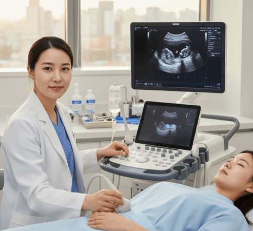
Sonohysterography, also known as saline infusion sonography (SIS), is a specialized type of ultrasound test that provides a detailed look at the inside of a woman’s uterus. The procedure involves gently filling the uterine cavity with sterile saline solution during a transvaginal ultrasound. The fluid creates contrast, making the inner lining of the uterus (endometrium) clearly visible on the ultrasound screen.
- 🔍 Purpose: To evaluate abnormalities in the uterus such as fibroids, polyps, scarring, or structural changes.
- 🧬 Method: Uses a combination of sterile saline and ultrasound imaging.
- 📊 Result: Offers more accurate imaging compared to a standard pelvic ultrasound.
This test is considered minimally invasive, safe, and highly effective for identifying uterine issues that may affect fertility or cause abnormal bleeding.
Why it’s done
Sonohysterography is often recommended when doctors suspect issues with the uterine cavity.
➡ Main reasons include:
- ✅ Abnormal uterine bleeding: Detecting the cause of heavy or irregular periods.
- ✅ Infertility evaluation: Checking if uterine abnormalities are interfering with conception.
- ✅ Recurrent miscarriages: Investigating if uterine scarring or shape abnormalities contribute to pregnancy loss.
- ✅ Suspected growths: Identifying polyps, fibroids, or endometrial thickening.
- ✅ Follow-up: Monitoring patients after surgery or medical treatment of uterine conditions.
Alternatives
Although sonohysterography is highly effective, there are several alternative diagnostic methods:
- 🧾 Transvaginal Ultrasound (TVUS)
➝ Standard imaging but sometimes limited in detail.
➝ May miss small polyps or subtle structural problems. - 🩻 Hysterosalpingography (HSG)
➝ Uses X-rays with contrast dye.
➝ Often used in infertility workups to check fallopian tubes and uterus. - 🔬 Hysteroscopy
➝ Involves inserting a small camera directly into the uterus.
➝ Allows direct visualization and treatment at the same time. - 🧬 MRI
➝ Provides excellent images of pelvic organs.
➝ Usually reserved for complex or unclear cases.
Note: Sonohysterography is often chosen because it combines clarity, safety, and affordability.
Preparation
Proper preparation ensures better results and comfort during the test.
- ⏩ Schedule: The procedure is usually done after menstruation but before ovulation (day 5–12 of the cycle).
- ⏩ Avoid pregnancy: The test is not performed if you may be pregnant.
- ⏩ Medication: Some women may be given antibiotics or pain relievers before the procedure.
- ⏩ Diet: No special fasting required, but you may be asked to empty your bladder before the test.
- ⏩ Inform doctor: If you have pelvic infections, allergies to latex, or previous gynecological surgeries.
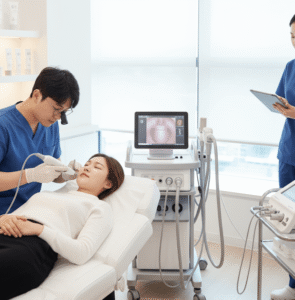
How it’s done
The procedure is simple, safe, and usually takes 15–30 minutes.
🩺 Step-by-step process:
- 👩⚕️ You lie on an exam table, similar to a Pap smear position.
- 👩⚕️ A speculum is gently inserted into the vagina to visualize the cervix.
- 👩⚕️ A thin catheter is inserted into the uterus through the cervix.
- 💧 Sterile saline solution is slowly infused into the uterus.
- 📡 A transvaginal ultrasound probe is used to capture images of the uterus while filled with fluid.
✔️ Mild cramping may occur as the saline is introduced.
✔️ No anesthesia is required.
✔️ The entire process is usually painless, though some women feel pressure or light discomfort.
Recovery
Recovery after sonohysterography is very quick, and most women return to normal activities the same day.
- 🔸 Mild spotting or watery discharge may occur for a few hours.
- 🔸 Some cramping may happen but usually subsides quickly.
- 🔸 Results are often available immediately as the doctor reviews the ultrasound images.
- 🔸 Follow-up may be scheduled if abnormalities are detected.
⚠️ Rarely, infection or injury can occur, but risks are minimal when performed by experienced doctors.
Treatment Options in Korea
South Korea is known for its advanced gynecological care, modern imaging technology, and affordable yet precise diagnostics.
➡ Why choose Korea for sonohysterography?
- 🏥 World-class hospitals with advanced ultrasound equipment.
- 👩⚕️ Expert gynecologists trained in reproductive medicine.
- 💳 Lower costs compared to the U.S. and Europe.
- 🌍 Medical tourism packages including interpretation and concierge care.
➡ If abnormalities are found during sonohysterography in Korea:
- Endometrial polyps → Removed via hysteroscopic surgery.
- Fibroids → Managed with medication, minimally invasive surgery, or laparoscopic procedures.
- Uterine adhesions → Treated with hysteroscopic adhesiolysis.
- Structural abnormalities (e.g., septum) → Surgically corrected for better fertility outcomes.
➡ Fertility-focused treatments:
Korea is a leader in IVF and reproductive medicine. Many women undergo sonohysterography as part of their fertility workup before assisted reproductive treatments.
Highlights of Sonohysterography in Korea
- ⭐ Safe, accurate, and minimally invasive diagnostic tool.
- ⭐ Performed by highly skilled gynecologists in modern clinics.
- ⭐ Quick results with advanced ultrasound imaging.
- ⭐ Affordable costs for both locals and international patients.
- ⭐ Integrated with fertility and women’s health programs.
✅ Summary: Sonohysterography in Korea provides a fast, safe, and reliable way to evaluate the uterine cavity. With Korea’s advanced medical technology, expert doctors, and patient-friendly care, women can access world-class diagnostic services at a fraction of the cost compared to many other countries.