What It Is
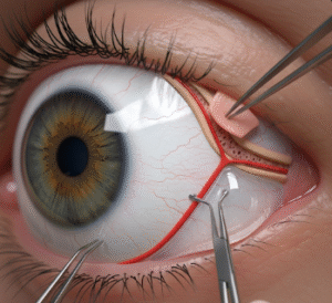
Canalicular laceration repair is a delicate microsurgical procedure used to repair injuries to the canaliculi, the small tear drainage channels in the upper and lower eyelids. These structures transport tears from the eye to the nasal cavity. Trauma from accidents, sharp objects, falls, or animal bites can cut the canaliculus, leading to constant tearing (epiphora), infection, and cosmetic deformity.
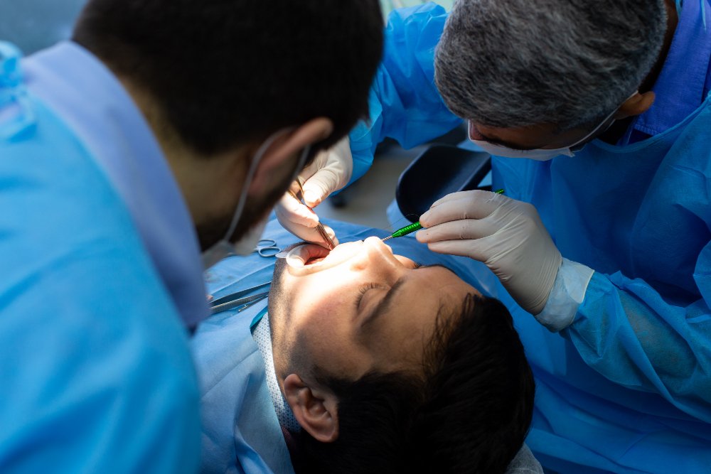
In Korea, repair is performed by oculoplastic surgeons using specialized instruments, fine sutures, and silicone stents to restore both eyelid appearance and tear drainage function.
Why It’s Done
Patients undergo canalicular laceration repair because:
- The injury causes persistent watery eyes (epiphora).
- Without repair, the canaliculus can scar shut, leading to permanent tearing.
- Eyelid shape and closure may be altered.
- Proper repair restores both function and aesthetics of the eyelid.
Good candidates include:
- Patients with a confirmed canalicular tear.
- Those in good general health who can tolerate local or general anesthesia.
- Patients seeking both functional tear drainage and cosmetic recovery.
Alternatives
- Observation: Only for very minor or superficial injuries not involving the canaliculus.
- Medical care: Antibiotics and wound care for infection prevention, but this does not restore drainage.
- No repair: Leads to permanent tearing and eyelid deformity.
Preparation
- Detailed consultation with an oculoplastic surgeon.
- Examination and probing of the lacrimal system to confirm the extent of damage.
- Early repair (within 24–48 hours) is often recommended for best results.
- Avoid smoking, alcohol, and blood-thinning medications before surgery.
How It’s Done
- Anesthesia: Local with sedation or general anesthesia depending on the patient and injury severity.
- Identification: The surgeon locates both cut ends of the canaliculus under magnification.
- Stenting: A silicone tube (monocanalicular or bicanalicular) is placed to keep the canaliculus open.
- Suturing: The canalicular tissue and eyelid layers are carefully sutured with fine stitches.
- Closure: Eyelid skin is closed with minimal scarring.
- Duration: 1–2 hours depending on complexity.
Recovery
- First week: Swelling, bruising, and discomfort are common.
- Skin sutures (if used) are removed in 5–7 days.
- Silicone stent remains in place for 3–6 months to ensure healing.
- Most patients resume light activities within a few days.
- Final results: Normal tear drainage and natural eyelid contour restored in several months.
Possible Complications
- Infection or irritation around the repair site.
- Stent displacement or loss.
- Persistent tearing if the repair does not heal properly.
- Visible scarring or eyelid deformity.
- Rare risks include canalicular blockage requiring revision surgery.
Treatment Options in Korea
Diagnosis
- Clinical examination and probing of the tear duct system.
- Digital imaging or CT if deeper trauma is suspected.
Medical Treatments
- Antibiotics for infection prevention.
- Lubricating eye drops to protect the surface.
Surgical or Advanced Therapies
- Monocanalicular stenting for simple tears.
- Bicanalicular intubation for more complex injuries.
- Microsurgical repair with sutures and stents for complete tears.
Rehabilitation and Support
- Regular follow-up visits to check stent position and healing.
- Use of lubricating drops and antibiotic ointments.
- Scar management if needed.
- Long-term monitoring of tear drainage function.
- International patients benefit from Korea’s specialized oculoplastic surgeons, advanced microsurgical equipment, and natural cosmetic outcomes.