What It Is
Orbital floor fracture repair is a surgical procedure to correct a fracture of the thin bone at the bottom of the eye socket (orbital floor). This type of injury often occurs after facial trauma, sports injuries, car accidents, or physical assault. When the orbital floor breaks, the eye can sink into the socket (enophthalmos), or soft tissues and muscles can become trapped, causing double vision, pain, or restricted eye movement.
The surgery involves repositioning displaced tissue and repairing the orbital floor using implants (titanium mesh, porous polyethylene, or absorbable materials) or bone grafts. Korean surgeons are known for using advanced 3D imaging and custom implants to achieve precise reconstruction and restore both appearance and function.
Why It’s Done
Patients undergo orbital floor fracture repair because:
- They have double vision (diplopia) or difficulty moving the eye.
- The eye has sunken into the socket (enophthalmos) or appears misaligned.
- They experience pain or numbness due to nerve compression.
- Large fractures threaten the function of the eye or long-term facial symmetry.
Good candidates include:
- Patients with moderate to severe fractures diagnosed on CT scans.
- Individuals with functional symptoms such as vision problems or cosmetic deformity.
- Healthy patients who can tolerate general anesthesia.
Alternatives
- Observation: Small fractures without symptoms may heal on their own with monitoring.
- Steroid or cold compress therapy: To manage swelling in mild cases.
- Non-surgical management: For patients with minimal symptoms and stable eye position.
Preparation
Before undergoing orbital floor fracture repair in Korea, patients will:
- Have a CT scan to evaluate the extent of fracture and soft tissue entrapment.
- Undergo ophthalmologic and surgical consultations.
- Avoid smoking and alcohol at least 2–4 weeks before surgery.
- Discontinue medications or supplements that increase bleeding.
- Prepare for a short hospital stay after surgery.
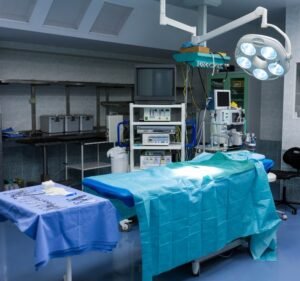
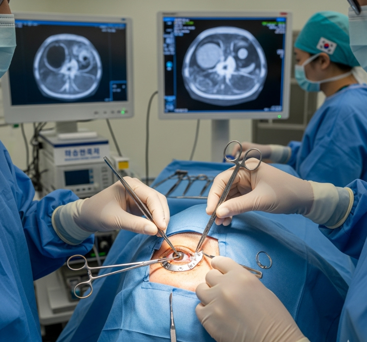
How It’s Done
- Anesthesia: General anesthesia is typically used.
- Incision: A small incision is made either inside the lower eyelid (transconjunctival) or just below the eyelashes, leaving minimal visible scarring.
- Tissue release: Trapped muscles and soft tissue are gently freed.
- Implant placement: The orbital floor is reconstructed using a titanium mesh, porous polyethylene implant, or resorbable plate. In some cases, bone grafts are used.
- Closure: The incision is carefully sutured.
- Duration: 1–2 hours, usually performed as an inpatient or day surgery.
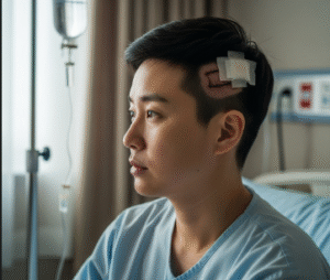
Recovery
- First week: Swelling, bruising, and mild pain around the eye are common. Cold compresses and medication help relieve discomfort.
- Vision: Temporary blurred or double vision may persist but usually improves as swelling resolves.
- Return to activities: Light activities in 1 week; strenuous exercise should be avoided for 4–6 weeks.
- Final results: Functional improvement (eye movement, vision) and cosmetic correction usually become clear within 2–3 months.
Possible Complications
- Persistent or recurrent double vision.
- Incomplete correction of enophthalmos (sunken eye).
- Infection or implant displacement.
- Scarring or eyelid malposition (rare with modern techniques).
- Rare risks: vision loss due to optic nerve damage.
Treatment Options in Korea
Diagnosis
Korean surgeons use high-resolution CT imaging, 3D facial scans, and ophthalmologic testing to evaluate the fracture and plan reconstruction.
Medical Treatments
For small, non-symptomatic fractures, observation, anti-inflammatory medications, and eye protection may be recommended.
Surgical or Advanced Therapies
- Traditional orbital floor reconstruction with titanium mesh or porous implants.
- Custom 3D-printed implants for precise anatomical repair.
- Endoscopic-assisted orbital floor repair, minimizing external scars.
- Combination with adjacent fracture repairs (zygomatic or maxillary fractures) if needed.
Rehabilitation and Support
- Postoperative monitoring with ophthalmologists to check vision and eye movement.
- Scar management (if external incision was made).
- Long-term follow-ups with CT scans to confirm implant stability.
- International patients benefit from multilingual care teams, advanced imaging, and modern implant technologies in Korean hospitals.