Overview
Ureterocele is a congenital condition in which the end of a ureter swells and balloons into the bladder, causing a cyst-like structure. This can obstruct urine flow, lead to kidney damage, and increase the risk of urinary tract infections. Ureteroceles are more commonly diagnosed in infants and children but can also be found in adults. Early detection and treatment help preserve kidney function and prevent complications.
What is Ureterocele
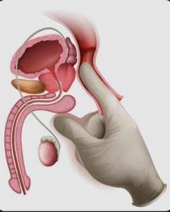
Ureterocele is a malformation of the distal ureter, where the ureter enters the bladder. Instead of forming a narrow, direct connection, the ureter balloons out, creating a sac-like pouch that may block the normal flow of urine. Ureteroceles can occur in one or both ureters and may be associated with duplicated collecting systems—a condition where a kidney has two ureters instead of one. The severity of symptoms depends on the degree of obstruction and whether one or both kidneys are affected.
Symptoms
Symptoms vary by age and severity of obstruction:
In infants and children:
- Recurrent urinary tract infections (UTIs)
- Fever
- Abdominal or flank pain
- Poor growth or feeding
- Blood in the urine (hematuria)
- Palpable abdominal mass (in some cases)
In adults:
- Frequent UTIs or painful urination
- Urinary incontinence or urgency
- Difficulty emptying the bladder
- Hematuria
- Flank pain or pressure
- Stones in the bladder or ureter
Causes
Ureterocele is a congenital condition, meaning it develops during fetal development. Causes include:
- Incomplete breakdown of the membrane at the ureteral orifice during development
- Abnormal ureteral development or insertion into the bladder
- Often associated with duplex collecting systems
- May occur without a known cause in otherwise healthy individuals
Risk Factors
- Female sex (more common in females)
- Congenital urinary tract abnormalities
- Family history of urologic malformations
- Duplicated ureter system
- Prematurity or low birth weight (increased risk of kidney/urinary tract anomalies)
- Obstructive uropathy detected on prenatal ultrasound
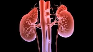
Complications
- Hydronephrosis (swelling of the kidney due to urine backup)
- Recurrent urinary tract infections
- Kidney damage or scarring
- Bladder outlet obstruction
- Vesicoureteral reflux (VUR) – backward flow of urine from bladder to kidney
- Bladder stones or urinary retention
- Loss of kidney function (in severe or untreated cases)
Prevention
Ureterocele cannot be prevented, as it results from congenital developmental abnormalities. However, early detection and appropriate intervention can minimize complications:
- Prenatal ultrasounds may detect hydronephrosis or ureterocele before birth
- Prompt evaluation of UTIs or voiding dysfunction in infants and children
- Regular urologic monitoring for individuals with known urinary tract anomalies
- Follow-up imaging to assess kidney function and structure
Treatment Options in Korea
South Korea offers advanced diagnostics and minimally invasive treatments for ureterocele in both children and adults:
- Ultrasound and voiding cystourethrogram (VCUG): To diagnose ureterocele and assess for reflux or obstruction
- Renal scan: To evaluate kidney function
- Endoscopic puncture (transurethral incision of the ureterocele): A minimally invasive first-line treatment to relieve obstruction and allow normal urine flow
- Ureteral reimplantation surgery: For persistent or complex cases, particularly in duplicated systems
- Partial nephrectomy: If a portion of the kidney is non-functional due to chronic obstruction
- Stent placement: Temporary relief of blockage in select cases
- Lifelong monitoring: For kidney function, recurrent infections, and urinary drainage
Korea’s pediatric and adult urology specialists use a tailored, patient-centered approach with modern surgical and endoscopic techniques to ensure optimal outcomes.