Overview
Arteriovenous Malformation (AVM) is a rare and potentially life-threatening condition involving an abnormal connection between arteries and veins, bypassing the capillary system. This disrupts normal blood flow and oxygen circulation and can lead to hemorrhage, seizures, or neurological damage, particularly when AVMs occur in the brain or spinal cord.
What is Arteriovenous Malformation?
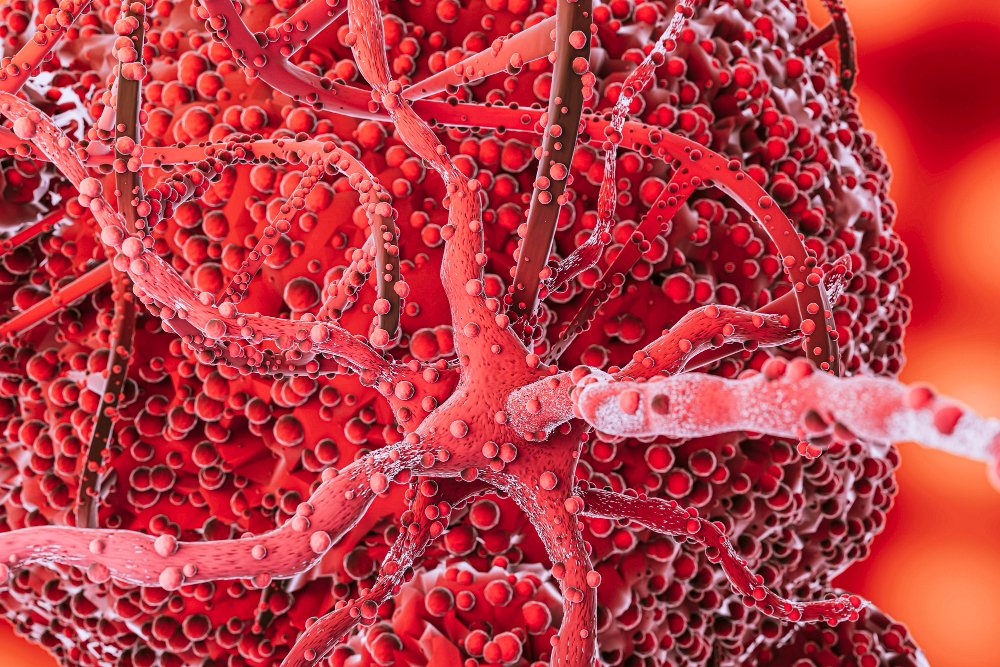
An AVM is a tangled cluster of blood vessels where arteries connect directly to veins. Normally, blood flows from arteries into capillaries, where oxygen and nutrients are exchanged with tissues, and then into veins. In AVM, this process is disrupted, often leading to high-pressure blood flow that can damage nearby tissues or cause vessels to rupture.
AVMs can develop anywhere in the body but are most dangerous when located in the brain, spinal cord, or gastrointestinal tract.
Symptoms
Brain AVM:
- Seizures
- Headaches
- Progressive neurological deficits (e.g., weakness, vision problems, numbness)
- Dizziness or difficulty speaking
- Sudden brain hemorrhage (stroke-like symptoms)
Spinal AVM:
- Back pain
- Muscle weakness
- Numbness or tingling
- Loss of bladder or bowel control
Other Locations (e.g., GI tract, limbs):
- Pain or swelling
- Skin discoloration
- Ulcers or bleeding
Causes
- Most AVMs are congenital, meaning they are present at birth, though their cause is often unknown
- Not usually hereditary
- Some AVMs may develop following trauma or as part of syndromes like Hereditary Hemorrhagic Telangiectasia (HHT)
Risk Factors
- Family history of AVM (rare)
- Presence of genetic syndromes such as HHT
- History of brain injury (in rare acquired cases)
- Being male (slightly higher incidence)
Complications
- Hemorrhage: Rupture of AVM vessels can cause life-threatening bleeding, especially in the brain
- Stroke or brain damage: Due to lack of oxygen in affected tissues
- Seizures and permanent neurological damage
- Hydrocephalus: If AVM obstructs cerebrospinal fluid flow
- Heart failure: In rare cases of very large AVMs causing significant blood shunting
Prevention
- There is no known way to prevent congenital AVMs
- Early diagnosis and monitoring can reduce complications
- Individuals with genetic syndromes should undergo regular screening
- Prompt medical attention for unexplained neurological symptoms
Treatment Options in Korea
South Korea offers cutting-edge care for AVMs through advanced imaging, neurosurgery, interventional radiology, and radiosurgery, especially at top-tier hospitals.
- Diagnosis
- MRI or CT Scan: Identifies AVM location and size
- Cerebral Angiography: Gold standard to map AVM blood vessels
- EEG: Used if seizures are present
- Spinal MRI: For AVMs located in the spine
- Monitoring
- Small, unruptured AVMs may be observed regularly
- Control of blood pressure and seizures through medication
- Medications
- Antiepileptic drugs (AEDs) for seizure control
- Pain management and anti-inflammatory medications as needed
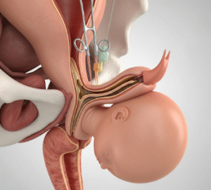
- Surgical Treatment
- Microsurgical Resection: Complete removal of AVM through open surgery; effective for accessible AVMs
- Endovascular Embolization: Minimally invasive; a catheter delivers glue-like substance to block abnormal vessels
- Stereotactic Radiosurgery (e.g., Gamma Knife): Precise radiation targets the AVM, causing gradual vessel closure over time
- Post-Treatment Care
- Regular follow-up imaging to ensure complete resolution
- Neurological rehabilitation for patients with post-operative deficits
- Lifestyle modifications and counseling