What it is
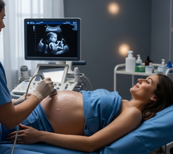
An ultrasound scan (also called sonography) is a non-invasive imaging technique that uses high-frequency sound waves to produce images of internal organs, tissues, and blood flow.
➡ Key facts:
- ✔ Uses sound waves rather than X-rays, making it safe and radiation-free
- ✔ Commonly used for pregnancy monitoring, abdominal, cardiac, and musculoskeletal examinations
- ✔ Available in hospitals, diagnostic imaging centers, and specialized ultrasound clinics across Korea
- ✔ Provides real-time imaging, aiding in diagnosis, treatment planning, and monitoring
💡 Ultrasound is safe for all age groups, including pregnant women and children, and offers immediate visualization of soft tissues.
Why it’s done
Ultrasound scans are performed to:
➤ Monitor pregnancy → Assess fetal growth, anatomy, and health
➤ Diagnose abdominal or pelvic conditions → Liver, gallbladder, kidneys, bladder, ovaries, or prostate
➤ Evaluate blood flow → Doppler ultrasound for arteries, veins, or heart function
➤ Guide procedures → Biopsies, drainages, or injections
➤ Assess musculoskeletal issues → Tendons, ligaments, muscles, and joints
⚠ Ultrasound helps detect diseases early, monitor ongoing conditions, and guide minimally invasive procedures safely.
Alternatives / Complementary Measures
Other imaging techniques include:
✔ X-ray → Better for bone imaging but uses radiation
✔ CT scan (Computed Tomography) → Detailed cross-sectional images; involves radiation
✔ MRI (Magnetic Resonance Imaging) → Detailed soft tissue imaging, no radiation
✔ Endoscopy → Direct visualization of internal organs
✔ Laboratory tests → Complement imaging for diagnosis
⚠ Ultrasound is preferred for soft tissue and real-time evaluation and is often combined with other imaging for comprehensive diagnosis.
Preparation
Preparation varies depending on the type of ultrasound:
🔹 Abdominal ultrasound → Fasting 6–8 hours to reduce gas and improve visualization
🔹 Pelvic ultrasound → Drink water to fill the bladder
🔹 Cardiac ultrasound (echocardiography) → Usually no fasting required
🔹 Medication → Usually no adjustment, but follow doctor’s instructions
🔹 Clothing → Wear loose clothing or bring a gown provided by the clinic
💡 Korean imaging centers provide specific pre-scan instructions to ensure accurate and high-quality imaging.
How it’s done
➡ Step-by-step ultrasound procedure:
- Positioning → Patient lies on an examination table
- Gel application → Conductive gel applied to the skin to improve sound wave transmission
- Probe placement → Transducer moved over the target area
- Real-time imaging → Technician or doctor captures images and evaluates structures
- Optional Doppler assessment → Measures blood flow and detects blockages or abnormalities
- Review and interpretation → Radiologist analyzes images and provides a report
💡 Ultrasound is painless, non-invasive, and usually takes 15–45 minutes, depending on the area being examined.
Effectiveness & Success Rate
✔ Highly effective for soft tissue and organ imaging
✔ Safe for repeated use, especially during pregnancy
✔ Real-time imaging allows immediate clinical decisions
✔ Combined with Doppler or contrast ultrasound, provides detailed vascular and organ assessment
💡 Korean hospitals use state-of-the-art ultrasound machines for high-resolution imaging and accurate diagnosis.
Recovery / Expected Outcomes
✔ Immediate recovery → Patients can resume normal activities immediately
✔ No anesthesia required → Minimal risk, no downtime
✔ Result interpretation → Radiologist report usually available same day or within 1–2 days
✔ Follow-up imaging → As needed for monitoring conditions or treatment effectiveness
💡 Ultrasound results help confirm diagnosis, guide treatment, and monitor disease progression safely.
Complications / Risks
⚠ Ultrasound is extremely safe, with negligible risks:
➡ Rare skin irritation from gel
➡ Minimal discomfort from probe pressure
➡ Inconclusive results → May require follow-up with CT or MRI for more detailed imaging
💡 Korean clinics prioritize patient safety and comfort, ensuring high-quality imaging with minimal risk.
Treatment Options in Korea (Post-Ultrasound Care)
🔹 Follow-up consultations → Discuss findings with doctor
🔹 Guided procedures → Ultrasound-guided biopsies, drainages, or injections
🔹 Referral to specialists → Based on imaging results (e.g., hepatologist, cardiologist, obstetrician)
🔹 Monitoring → Regular ultrasound follow-ups for chronic conditions or pregnancy
🔹 Preventive measures → Lifestyle or medical advice depending on diagnosis
💡 Ultrasound in Korea is integrated into diagnostic, treatment, and monitoring pathways, ensuring seamless patient care.
Top Hospitals & Clinics in Korea for Ultrasound
🏥 Seoul National University Hospital (SNUH) – Advanced imaging for multiple specialties
🏥 Asan Medical Center (Seoul) – High-resolution ultrasound and Doppler imaging
🏥 Samsung Medical Center (Seoul) – Specialized obstetric and abdominal ultrasound services
🏥 Yonsei Severance Hospital – Pediatric and cardiac ultrasound expertise
🏥 Private diagnostic centers nationwide – Accessible, rapid service, and modern equipment
Conclusion
Ultrasound scanning in Korea is a safe, effective, and essential imaging tool for diagnosis, treatment guidance, and monitoring of a wide range of medical conditions.
✔ Non-invasive, radiation-free, and suitable for all ages
✔ Provides real-time images for accurate assessment of organs, blood flow, and fetal health
✔ Korean hospitals ensure advanced technology, expert radiologists, and timely reporting
✔ Enables early detection, treatment planning, and ongoing monitoring
By integrating state-of-the-art ultrasound technology with professional interpretation and follow-up care, Korea provides high-quality diagnostic imaging that supports optimal health outcomes.